
What percentage of indeterminate calcifications are malignant?
By Robert Guerrero
What percentage of indeterminate calcifications are malignant?
Almost half (47.7%) of the cases were graded as indeterminate, 28.3% cases as suspicious and 24.0% cases as highly suspicious. After assessment, 47.9% of cases were malignant, 4.8% cases were indeterminate (including atypia) and 47.3% cases were benign.
What is tomosynthesis guided biopsy?
Tomosynthesis-guided core biopsy facilitates tissue diagnosis for mammographic abnormalities seen on 2D and 3D mammograms, including asymmetries, focal asymmetries, masses, architectural distortions, and calcifications.
What is tomosynthesis diagnostic?
Tomosynthesis is an imaging, or X-ray, technique that can be used to screen for early signs of breast cancer in people with no symptoms. This type of imaging can also be used as a diagnostic tool for people who are having breast cancer symptoms. Tomosynthesis is an advanced type of mammography.
What is the meaning of tomosynthesis?
Tomosynthesis or “3D” mammography is a new type of digital x-ray mammogram which creates 2D and 3D-like pictures of the breasts. This tool improves the ability of mammography to detect early breast cancers, and decreases the number of women “called back” for additional tests for findings that are not cancers.
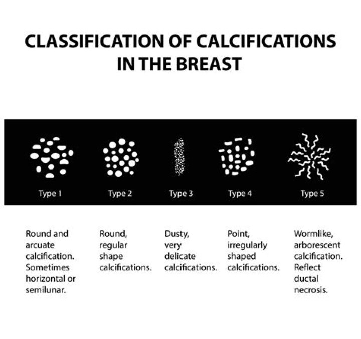
What are indeterminate calcifications?
Indeterminate calcifications: Any calcifications that do not clearly fit into any of the categories above. The radiologist will decide if a short-term follow-up mammogram is indicated (within four to six months) to ensure calcifications are stable.
What are heterogeneous calcifications?
Coarse heterogeneous calcifications are irregular calcifications that are between 0.5 mm and 1 mm in size. They are usually in a group and are smaller in size than dystrophic calcifications.
Can you take a shower after a breast biopsy?
You can shower 24 hours (1 day) after your biopsy. Remove your bandage before showering but leave the Steri-Strips in place. Let the shower water run over your biopsy site. Gently pat it dry with a clean towel.
What type of imaging uses low level radionuclides to locate and stage tumors?
Nuclear scans make pictures based on the body’s chemistry (like metabolism) rather than on physical shapes and forms (as is the case with other imaging tests). These scans use liquid substances called radionuclides (also called tracers or radiopharmaceuticals) that release low levels of radiation.
Is tomosynthesis the same as ultrasound?
Prof Houssami said: “In this study we are comparing two additional tests to see if they can do better than standard mammograms in finding cancer in women with dense breasts; we have found that ultrasound does better than tomosynthesis, but ultrasound is a separate test, it is time-consuming and, in less experienced …
Is tomosynthesis better for dense breasts?
No improvements were seen in decreased recall visits scheduled or a higher cancer detection rate.
Is tomosynthesis an ultrasound?
Screening with tomosynthesis or ultrasound detects more cancers in dense breasts. Summary: In women with dense breasts, adding either tomosynthesis (a form of 3-D mammography) or ultrasound scans to standard mammograms can detect breast cancers that would have been missed, according to new results.
What is a Tomo 3D mammogram?
Overview. A 3D mammogram (breast tomosynthesis) is an imaging test that combines multiple breast X-rays to create a three-dimensional picture of the breast. A 3D mammogram is used to look for breast cancer in people who have no signs or symptoms.
What is tomosynthesis (3D mammography)?
What Is Tomosynthesis (3D mammography)? Tomosynthesis or “3D” mammography is a new type of digital x-ray mammogram which creates 2D and 3D-like pictures of the breasts. This tool improves the ability of mammography to detect early breast cancers, and decreases the number of women “called back” for additional tests for findings that are not cancers.
Can tomosynthesis be used for routine screening?
Tomosynthesis has been approved for use in the NHS breast screening programme ( BSP) as an optional extra tool in the assessment of screen detected soft tissue breast abnormalities. It must not be used for routine screening outside of a clinical trial approved by the breast screening research advisory committee ( RAC).
Can tomosynthesis be used to diagnose multifocal breast cancer?
Unexpected multifocal disease may be found on tomosynthesis images. A full assessment is needed for any additional incidental lesions identified on tomosynthesis as set out in the clinical guidance for breast screening assessment. Studies have found that tomosynthesis can aid interpretation in dense breasts.
When is tomosynthesis indicated in the work up of soft tissue masses?
If additional mammography is required in the work up of masses and asymmetric densities, tomosynthesis is suggested in the first instance to: Ill-defined soft tissue masses without architectural distortion or microcalcification may be very inconspicuous on tomosynthesis.