What are the types of autoradiography?
By Olivia House
What are the types of autoradiography?
The following three types of radiations are used in autoradiography: Alpha rays – The alpha rays particles which consist of 2 neutrons and 2 protons and infact charged helium atoms. Radium 226 is their source. Beta rays – The beta rays are electrons ejected or emitted by nuclei.
What is the concept of autoradiography?
Autoradiography: A technique using X- ray film to visualize molecules or fragments of molecules that have been radioactively labeled. Autoradiography can, for example, be used to analyze the length and number of DNA fragments after they are separated from one another by a method called gel electrophoresis.
How was autoradiography discovered?
Autoradiography was discovered at the beginning of last century when Henri Becquerel observed that a “mysterious” radiation coming from uranium impressed photographic plates (Becquerel, 1896). This radiation was later given the name of gamma radiation by Rutherford (1903).
What are the techniques of autoradiography?
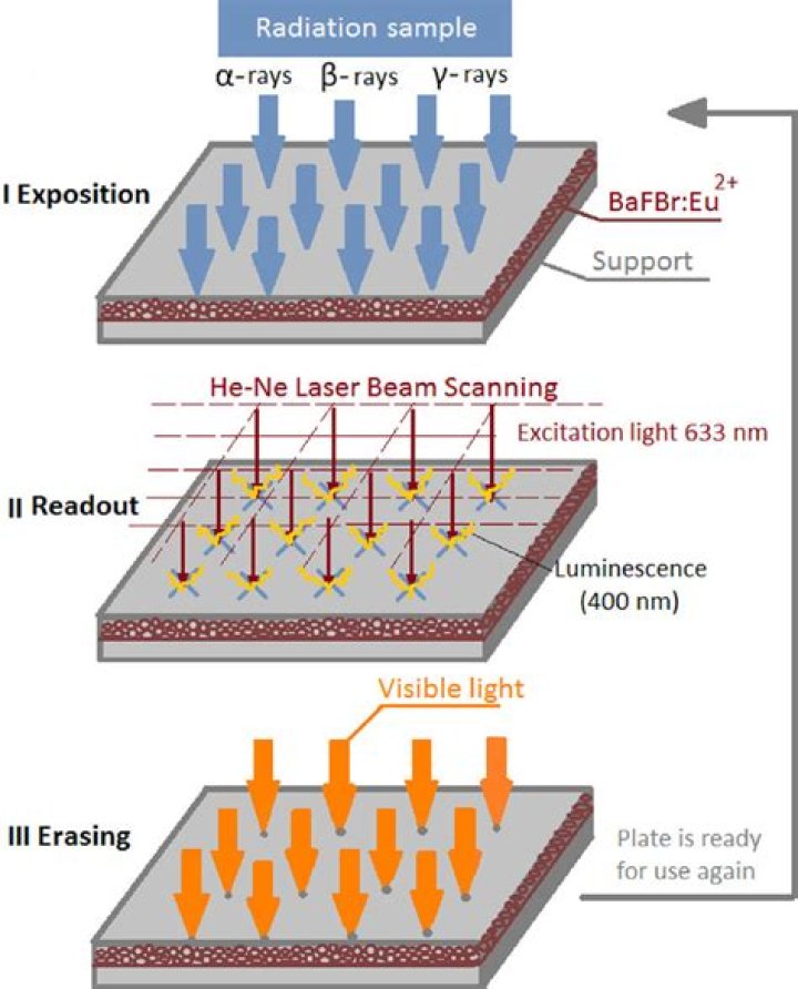
Autoradiography is a technique using X- ray film, phosphor imaging plates, beta imaging systems, or photo-nuclear emulsion to visualize molecules or fragments of molecules that have been radioactively labeled, and it has been used to quantify and localize drugs in tissues and cells for decades.
What is quantitative autoradiography?
Quantitative autoradiography of macroscopic specimens using computer-assisted image analysis is now widely used for studying the distribution of peptide receptors in the brain and peripheral tissues and more recently has been used to measure mRNA in tissue sections by in situ hybridisation.
Is autoradiography still used?
In vivo autoradiography using laboratory animals is widely used in metabolic studies, disease monitoring and new drug development experiments….Autoradiography In The Clinical Context.
| Positron Isotope | Half-life (min) | Application |
|---|---|---|
| 68-gallium (68Ga) | 68.3 | Detection of neuroendocrine tumors |
Why gelatin is used in autoradiography?
General Principle Of Autoradiography Each AgX molecule is individually encapsulated in the gelatin, and functions as an independent detector of radioactive decay from the radiolabeled sample. Once radioactive particles hit the gelatin emulsion, AgX is reduced resulting in the production of insoluble silver crystals.
Who invented autoradiography?
In 1867, Niepse de Saint Victor first described the phenomenon of autoradiography, which he described as the “persistent activity due to an unknown chemical radiation” (3).
Which emitters are suitable for autoradiography?
Weak b-emitter such as 3H, 14C and 35S which are suitable for autoradiography .
What is autoradiography in biology?
Autoradiography records the distribution of radioactive materials in botanical and histological specimens placed in contact with a photographic emulsion. This technique has been applied to the study of metabolism of plants and animals; it records the activity of organic compounds of radioactive isotopes introduced…
Why is it called autoradiography?
An autoradiograph is an image on an x-ray film or nuclear emulsion produced by the pattern of decay emissions (e.g., beta particles or gamma rays) from a distribution of a radioactive substance. The film or emulsion is apposed to the labeled tissue section to obtain the autoradiograph (also called an autoradiogram).
What are the most commonly used isotopes in autoradiography?
Common radioisotopes in autoradiography are sulfur-35, hydrogen-3, carbon-14, 125-iodine or phosphorus-32 (35S, 3H, 14C, 125I and 32P, respectively) which are used to determine the distribution of the radiolabeled molecules in tissues, cells or cellular organelles (Figure 1), but also in the study of protein …
What is autoradiography and when was it discovered?
Autoradiography was discovered at the beginning of last century when Henri Becquerel observed that a “mysterious” radiation coming from uranium impressed photographic plates (Becquerel, 1896 ).
What is GTP autoradiography used for?
[35S]GTPγS autoradiography has been used to identify receptor-activated G proteins for several receptor systems (see Table I). [35S]GTPγS autoradiography has been used in several species, including rat, mouse, guinea pig, chick, and monkey.
What is the resolution of autoradiography?
Depending on the use of absorbers, autoradiography can be sensitive to alpha particles, beta particles, or both, but the resolution is typically around 50 μm (see Fig. 13.11 ). Figure 13.11. Cropped autoradiographs of two sawed low-enriched UO 2 subaliquots, which were mounted in epoxy and polished before being exposed to an SR IP for 25 h.
What are the applications of autoradiography in biochemistry?
The autoradiography can be employed in different studies, among them, those concerning visualizing and quantitating radiolabeled proteins or DNA separated by PAGE, as well as image analysis for quantitating radiolabeled protein drug concentration in serum for pharmacokinetic studies.